Allergy
Blood Diseases
Bone & Joints
Brain
Cancer
Child Care
Cosmetic Surgery
Diabetes
Endocrinology
ENT
Eye
Gen Medicine
General Surgery
Heart
Kidney
Lifestyle
Liver & Digestive
Lung
Men’s Health
Mental health
Physiotherapy
Rheumatology
Skin and hair
Sleep Disorders
Spine
Transplant
Women Health
Thyroid
Vascular Surgery
Cerebral Aneurysm

Neuro Surgeon
Hyderabad | 06 Nov 2023
What is a Cerebral Aneurysm?
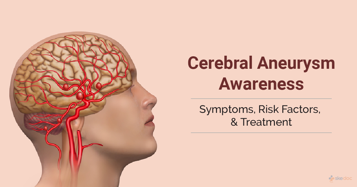
A Cerebral Aneurysm, also known as Brain aneurysm, is a condition in which a defect or weakness in the walls of the blood vessels of the brain results in localized swelling, dilation, or ballooning of these vessels.
Alternate name
Intracranial Aneurysm
Is cerebral aneurysm a medical emergency?
A cerebral aneurysm can rupture or leak and can lead to conditions such as hemorrhagic stroke or subarachnoid haemorrhages (SAH), which are medical emergencies.
Types
It can be classified based on their size as:
- Small - Less than 15 mm
- Large - 15 to 25 mm
- Giant - 25 to 50 mm
- Supergiant - More than 50 mm
They can also be of the following types:
- Saccular Aneurysm or Berry Aneurysm: They are the most common type of cerebral aneurysms and appear like a berry on a stem
- Fusiform Aneurysm: The dilation or ballooning of the vessel wall happens all around and is not limited to one side. Usually associated with atherosclerosis.
- Dissecting Aneurysm: A tear in the layers of the blood vessel and leakage of blood into the gap between the layers results in a bulging or dilation of the vessel wall
- Microaneurysms or Charcot Bouchard Aneurysms: It is usually seen in the small blood vessels and is associated with chronic hypertension
Causes
The cause of a cerebral aneurysm is not fully understood, but multiple factors contribute to its formation. Structural defects of the walls of the blood vessels, which may be either acquired or inborn, can result in the formation of an aneurysm. The gradual degeneration of the vessel wall, followed by its weakening and loss of elasticity, and the pressure from the blood flowing through it results in abnormal dilation and ballooning. It is also more commonly seen where the vessel bifurcates to create smaller branches.
Risk factors
The risk factors for the development of a cerebral aneurysm may include the following:
- Acquired risk factors:
- Age - The risks increase with increasing age.
- Alcohol consumption
- High blood pressure
- Cigarette Smoking
- Trauma to the head
- High cholesterol levels and atherosclerosis.
- Infections - Especially fungal
- Cancers
- Use of recreational drugs such as cocaine.
- Inherited risk factors:
- Gender - Women are at a greater risk
- Family history of aneurysms
- Inherited connective tissue disorders - Ehler-Danlos syndrome, Fibromuscular dysplasia, Marfan's syndrome
- Coarctation of the aorta - Abnormally narrow aortae
- Arteriovenous malformations in the brain
- Sickle cell anemia
- Polycystic kidney disease
- Neurofibromatosis
- Tuberous sclerosis
Signs & symptoms
A majority of cerebral aneurysms may not show any symptoms until they rupture, while some of them may show symptoms due to the pressure effects on adjacent structures.
The symptoms and signs may include the following:
An unruptured aneurysm may present with:
- Headache - Not very common if unruptured
- Eye pain, eye movement difficulties, dilated pupil, and visual disturbances
- Numbness on one side of the face
A ruptured aneurysm may present with:
- Rapid onset of intense and severe headache
- Stiff neck
- Nausea and vomiting
- Pain in the eyes, dilated pupils, blurring of vision, or double vision
- Sensitivity to light or sound
- Drowsiness or loss of consciousness
- Loss of balance and coordination
- Seizures
- Breathing difficulties
- High blood pressure
- Bleeding from the nose
Investigations
The following investigations may be done:
- Laboratory tests:
- CBP & ESR
- Prothrombin time
- Liver function tests
- Serum electrolytes
- Arterial blood gases
- Lumbar puncture: To evaluate the cerebrospinal fluid for the presence of blood in the event of a rupture.
- Imaging tests:
- CT scan: CT angiography was done without a contrast dye as the dye can mask the presence of a subarachnoid hemorrhage.
- MRI: MRI angiography
- Cerebral angiography
- ECG
- EEG
Diagnosis
A diagnosis of a cerebral aneurysm is established based on medical history, clinical evaluation, and the results of the investigations done.
Course & Stages
A cerebral aneurysm may be graded as follows:
- Grade 0: Unruptured aneurysm
- Grade 1: No symptoms or minimal headache, slight neck stiffness, no blood on CT
- Grade 2: Moderate to severe headache and neck stiffness, cranial nerve involvement, a diffuse thin layer of blood on CT.
- Grade 3: Drowsiness and confusion, and a thick layer of blood on CT.
- Grade 4: Extreme drowsiness, moderate to severe weakness on one side of the body, intracerebral or intraventricular bleeding seen on CT.
- Grade 5: Deep coma, decerebrate posturing (hands turned inwards, toes pointing downwards, head and neck arched backwards, and muscles are tight and rigid).
Treatment options
The treatment of cerebral aneurysms that have ruptured includes surgical interventions and supportive medical management.
Medical management
The medical management of a ruptured cerebral aneurysm may include:
- Calcium Channel blockers: To improve the neurological deficits due to SAH, should be started within 96 hours after an SAH
- Antiepileptics: To prevent and treat seizures
- Antihypertensives: To control the blood pressure
- Analgesics
- Anti-emetics
- Antacids
- Stool Softeners
Interventional including surgery and indications for surgery
Surgical management is done for ruptured aneurysms or aneurysms that have a very high risk of rupture.
The surgical procedures that are done to manage a cerebral aneurysm include:
- Open craniotomy and surgical clipping: The procedure is done by removing a part of the skull, identifying and locating the aneurysm, clipping the neck of the aneurysm to prevent blood from flowing into the dilated part of the vessel, and then replacing the removed piece of the skull.
- Endovascular coiling or coil embolization: It is a minimally invasive procedure that is done using image guidance and a catheter introduced through an artery in the groin to place tiny platinum coils inside the aneurysm, which results in the aneurysm from being blocked and thereby prevents the risk of rupture.
Other interventional procedures that are done include:
- Ventricular and lumbar draining catheters: To reduce the pressure inside the skull
- Shunt surgery: A shunt may be placed starting in the brain and ending in the abdomen to relieve pressure inside the skull
Role of Diet/ Exercise/ Lifestyle changes/ Preventive measures
Some measures that can be taken to lower the risk of rupture when a cerebral aneurysm is present include:
- Cessation of smoking
- Avoiding alcohol consumption
- Keeping blood pressure and blood cholesterol under control
- Eating a healthy diet and getting regular exercise are permissible
Individuals who have suffered a rupture of a Cerebral Aneurysm may need the following:
- Physical rehabilitative therapy
- Speech rehabilitative therapy
- Occupational rehabilitative therapy
Complications
The complications of a Cerebral Aneurysm and its rupture may include the following:
- Repeated bleeding
- Spasm of the blood vessels supplying to other parts of the brain leading to ischemic stroke
- Seizures
- Hydrocephalus - This may be caused due to obstruction to the outflow of the cerebrospinal fluid by the leaked blood from the ruptured aneurysm.
- Abnormal heart rhythms, heart attack, and heart failure
- Lung edema, pneumonia, and lung collapse
- Gastrointestinal bleeding
- Anemia
- Low sodium levels in the blood lead to permanent brain damage
Prognosis
The prognosis for a cerebral aneurysm and a ruptured cerebral aneurysm that results in a subarachnoid hemorrhage depends on the following factors:
- Age of the individual
- The neurological status at the time of hospital admission
- Location of the aneurysm
- The time delay between the aneurysm rupture and SAH and admission into the hospital
- Presence of high blood pressure and other illnesses
- The grade of the SAH
When to contact the doctor or hospital? / How to identify the emergency or complications?
It is advisable to seek immediate medical attention in the presence of an intense, severe headache accompanied by any other symptoms of a ruptured cerebral aneurysm.
Indications for hospitalization if required
Hospitalization will be required for the management of a cerebral aneurysm that has ruptured.
Screening methods
Screening for cerebral aneurysm via imaging tests is recommended for the following individuals:
- A family history of cerebral aneurysms.
- Inherited disorders and medical conditions pose an increased risk of developing cerebral aneurysms.
Suggested clinical specialist/ Departments to consult for this condition
Specialists from the Emergency Department and the Department of Neurology will attend a Cerebral Aneurysm.
Share this:
Was this article helpful?
YesNo
Comments

Ask a
Question!
Popular Searches
skedoc brings you healthcare that is relevant to your specific health needs. We make finding the Right Doctor and the Right Advice extremely easy.
skedoc brings you healthcare that is relevant to your specific health needs. We make finding the Right Doctor and the Right Advice extremely easy.
West Bengal
See More On Skedoc
Want to be notified when our article is published? Enter your email address and name below to be the first to know
 Sign in with Google
  Sign in with Facebook
Already have an account? Login here